Главная страница Случайная страница
Разделы сайта
АвтомобилиАстрономияБиологияГеографияДом и садДругие языкиДругоеИнформатикаИсторияКультураЛитератураЛогикаМатематикаМедицинаМеталлургияМеханикаОбразованиеОхрана трудаПедагогикаПолитикаПравоПсихологияРелигияРиторикаСоциологияСпортСтроительствоТехнологияТуризмФизикаФилософияФинансыХимияЧерчениеЭкологияЭкономикаЭлектроника
Echocardiography can be used to reveal vegetation on the cusps of the aortic valve, and less frequently on the mitral valve.
|
|
Treatment. The patient should be hospitalized and given antibiotics in large doses.
Heart Valvular Diseases
Stable pathological changes in the structure of the heart that interfere with its normal function are called heart disease. Congenital and acquired diseases of the heart are distinguished. The incidence of acquired heart diseases is much higher.
Congenital diseases of the heart arise due to abnormal development of the heart and the great vessels during the intrauterine growth of the foetus with preservation of the intrauterine character of circulation after birth. In defective division of the primary single-chamber arterial trunk into the pulmonary trunk and the aorta, and during formation of the heart chambers, defects in the interatrial and interventricular septa may be formed along with various abnormalities in the arrangement of the great vessels and their narrowing. Preservation of the intrauterine character of circulation after birth is the cause of patent ductus arteriosus (Botallo's duct) and patent foramen ovale. Congenital heart defects may often combine with communicated greater and lesser circulation systems and stenosis of the great vessels. Moreover, the valves (bicuspid, tricuspid, aortic, and pulmonary valves) may also have congenital defects.
Endocarditis, and especially rheumatic endocarditis, is the main cause of acquired heart defects. Less frequently heart disease is the result of sep sis, atherosclerosis, syphilis, injuries, etc. Inflammatory processes occurr ing in the valve cusps often end in their sclerosis: deformation and shorten ing. An affected valve does not close completely to cause valvular in competence. The cusps of the valves may adhere to one another because of inflammation to narrow the orifice they close. This narrowing is called stenosis.
MITRAL INCOMPETENCE
Incompetence of the mitral (bicuspid) valve (mitral insufficiency) is incomplete closure of the atrioventricular orifice during left-ventricular systole. As a result, the blood is regurgitated from the ventricle back to the atrium. Mitral incompetence may be organic and functional.
Organic insufficiency arises as a result of rheumatic endocarditis. Connective tissue develops in the cusps of the mitral valve which then contracts
To shorten the cusps and the tendons. The edges of the affected valve do not meet during systole and part of the blood is regurgitated through the slit into the left atrium from the ventricle during its contraction.
In functional (relative) incompetence the mitral valve is not altered but the orifice, which it has to close, is enlarged and the cusps fail to close it completely. Functional incompetence of the mitral valve may develop because of dilatation of the left ventricle (in myocarditis, myocardial dystrophy, or cardiosclerosis) and weakening of the circular muscle fibres that form the ring round the atrioventricular orifice. Affection of papillary muscles may also cause functional mitral incompetence. Functional insufficiency thus depends on dysfunction of the muscles responsible for the closure of the valve.
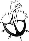
Haemodynamics. If the mitral valve fails to close completely during systole of the left ventricle, part of the blood is regurgitated into the left atrium. Blood filling of the atrium thus increases (because of the blood from the pulmonary veins which is added to the normal blood volurne). Pressure in the left atrium increases, the atrium is dilated and becomes hypertrophied.
The amount of blood that is delivered into the left ventricle from the overfilled left atrium during diastole exceeds normal and the atrium is thus overfilled and distended. The left ventricle has to perform excess work and becomes hypertrophied. Intensified work of the left ventricle compensates for the mitral incompetence during a long time (Fig. 69). When the contractile power of the left ventricular myocardium weakens, diastolic pressure in it increases and this in turn increases pressure in the left atrium.
|
|
|